Those of you who grew up in the 1980s might remember Billy Idol’s rock ballad “Eyes Without a Face.” It’s a catchy little song that owes its title to Jean Redon’s grisly horror novel Les yeux sans visage, which was adapted to the big screen back in 1962 and featured serial murders, a doctor’s daughter in an eerie face mask, and lots of gore, although the film version apparently toned it down quite a bit. Billy Idol’s sung version cast off all the gore and just went for despair and angst with strong guitars and a catchy rock beat. Nevertheless, if you’ve heard the song, you probably remember it, in part because of the simplicity of the title (and the awesome backup vocals by Perri Lister, in French and English).
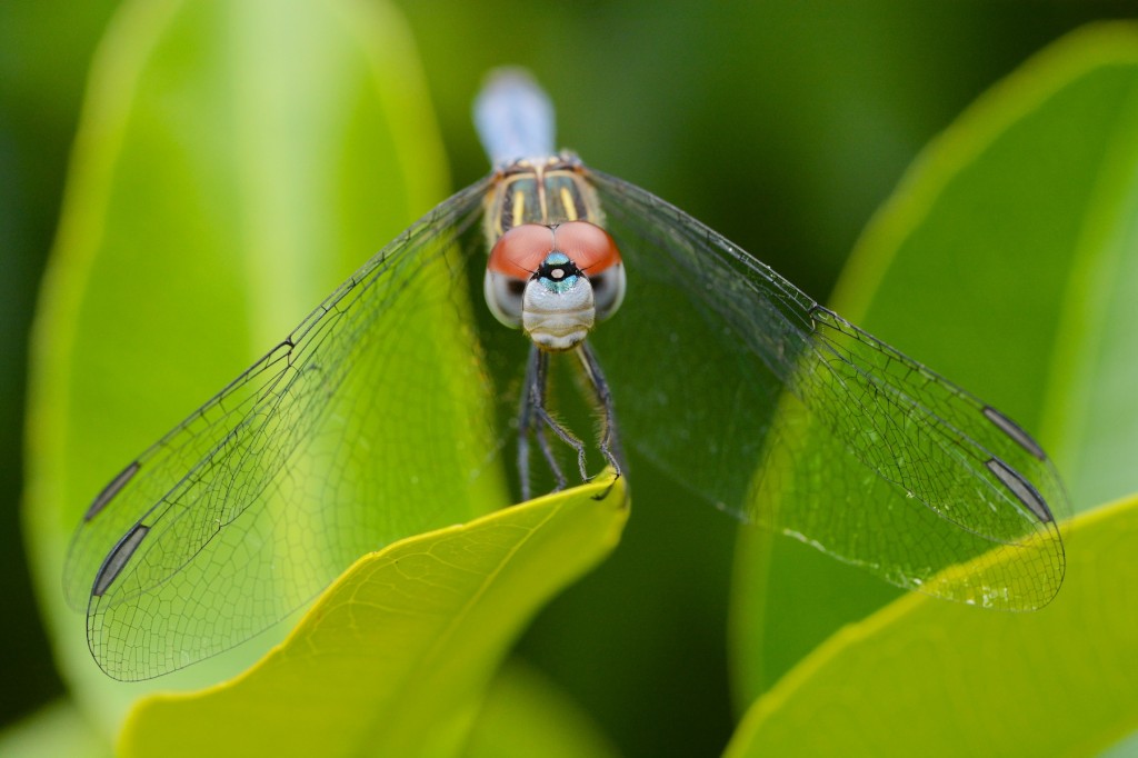
A lot of you might think that the dragonfly, with its enormous eyes, can also be described by that pithy little phrase. After all, their eyes are enormous, taking up most of their head. But, as you can see below, while it might make for a nice catchy song, it doesn’t describe the dragonfly very well. Dragonflies definitely have a “face.”

The dragonfly in question is a Blue Dasher (Pachydiplax longipennis), one of the most widespread and abundant dragonflies in North America. Apparently its larvae are able to tolerate a wide range of water quality, and compete in both fishful and fishfree ponds, which gives them a huge range of habitat in which to reproduce. They also make wonderful photographic subjects, often sitting nice and still to let the careful photographer get as close as his or her equipment will allow. (If you have a macro lens, that’s pretty darn close. Close enough to be able to crop an image and give you a little lesson in dragonfly facial anatomy.)
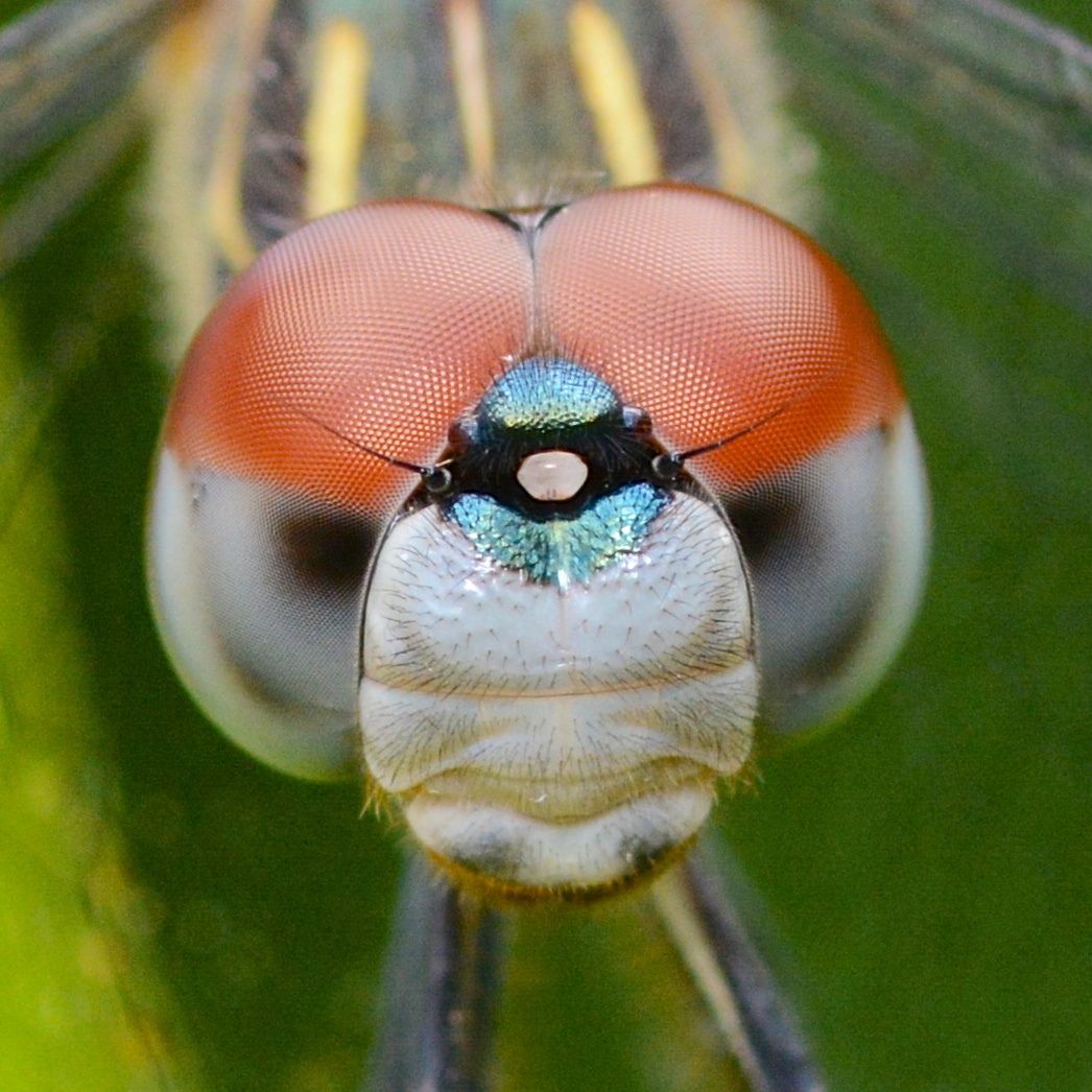
This picture was taken in my normal study site, aka my backyard, from about a foot and a half away; when I tried to pull in even tighter, the dasher lived up to its name and dashed away (flying of course; dragonflies, despite having six legs like most other insects, can’t walk). But when your camera has way too many megapixels (actually, that hardly matters at all) and you’re able to hold it nice and steady to get a sharp image (that’s the important bit), you can blow up a crop to just about whatever size you please. So I decided to spend a few minutes in Photoshop to give you all a little roadmap of the dragonfly’s facial anatomy.
Hover your cursor over the image to see what each structure is.

The “face” of the dragonfly, starting from the very bottom, consists of mandibles (not labeled, barely visible as dark smudges at bottom) that are usually concealed under the labrum, or “lower lip” (labeled). The clypeus is composed of the “top lip,” or postclypeus, and the anteclypeus, which is in what would be the “tongue” position were this a human face. Above the clypeus is the frons, from which arises the vertex, which serves as the anchor for the three ocelli, or simple eyes. The antennas arise from the base of the vertex as well. At the top of the “face,”the two eyes meet; the “joint” or seam is called the occiput.
Now that we’ve got the unfamiliar parts of the dragonfly face out of the way, let’s go back to that most familiar, and most prominent, feature of the dragonfly head: the huge compound eyes, with thousands upon thousands of ommatidia, or facets, that make up this complex visual apparatus. If you look closely, you’ll see that the larger facets are all on the top of the eye, the part that’s colored deep red. The smaller facets are on the bottom half, the part that’s colored blue-gray and has the black “pseudopupils.”
Most dragonflies that you’re likely to see fly during the day (the twilight-flying dragonflies are quite a bit less common in most people’s experience) and have excellent color vision. Our own eyes have three different color-sensitive retinal proteins, called opsins, that give us the familiar red-green-blue mix of color perception. (Those RGB color values don’t correspond exactly to the peak sensitivity of each opsin, but, meh… close enough). Diurnal (day-flying) dragonflies, on the other hand, have as many as five opsins in their visual reception apparatus, giving them access to a much greater range of the electromagnetic spectrum.
What’s interesting is the arrangement of the ommatidia with respect to the part of the spectrum they’re sensitive to. Recall the visual spectrum mnemonic: ROYGBV. That is, red-orange-yellow-green-blue, in ascending wavelength order. Red has the longest wavelength, while blue, violet, and, beyond the violet, ultra-violet, have the shortest wavelengths:
Those larger facets on the top of the dragonfly’s eye? They are sensitive to UV light and blue light (on the right in the image above). The smaller ommatidia on the bottom half of the eye? They’re specialized for longer-wavelength light, like orange or green.
As anyone who’s tried to get close to a dragonfly, whether to capture it or just its image, those eyes are extraordinarily capable of detecting movement. That great visual acuity appears to be related to the presence (and direction) of the pseudopupil (the dark region in the light blue-gray portion of the eye in the photo above). They seem to indicate the insect’s fovea, or zone of greatest visual acuity.
Land and Nilsson (2012) write that “perhaps the most useful feature of the pseudopupil [to the entomologist, if not to the insect itself] is that one can use it to measure inter-ommatidial angles. If one rotates an insect’s head through a degrees, and the pseudopupil appears to move across b facets, then the inter-ommatidial angle is a/b degrees. Variations in inter-ommatidial angle in different planes, and in different regions of the eye, can be mapped in this way, revealing how the eye is is organized to make the most of its limited visual acuity.”
Mapping the organization of the dragonfly eye is exactly what Truman Sherk did in a classic series of articles in the mid-1970s, tracing the development of dragonfly eyes from larva through adult animal, mapping the foveae and relating it to predation style. In the photo above you can see the pseudopupil looking directly at the camera, “anteriorly” as the scientists like to say. That is, directly in front of the animal.
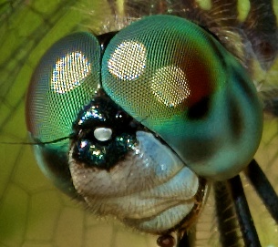
In the example below, though, you can see lots of “accessory” pseudopupils:

That is, in addition to the main, large pseudopupil, you can see several other dark spots; each of these is also a pseudopupil and indicates a zone of great visual acuity. According to Sherk, “the size and shape of the accessory pseudopupils [appear] to be related to the geometrical arrangement of the corneal facets, the slight curvature of the individual facets, and to the regional radius of curvature of the cornea. The contrast between the accessory pseudopupils and the surface [appears] to be related to the maturity of the cells between the cornea and the rhabdom.”
That might be a bit more than you wanted to know about the dragonfly and its face, but next time you see a dragonfly buzzing off when you so much as twitch a muscle in its direction, you might have a deeper appreciation for what makes that response possible: those amazing eyes.
References
Corbet, P. (1999). Dragonflies: Behavior and Ecology of Odonata. Ithaca: Cornell UP
Land, M. F., and D.-E. Nilsson. (2012). Animal Eyes, 2d ed. New York: Oxford UP.
Needham, J. G., and M. J. Westfall. (1955). A Manual of the Dragonflies of North America (Anisoptera). Berkeley: U of California P.
Paulson, D.R. (2011). Dragonflies and Damselflies of the East. Princeton, NJ: Princeton UP.
Sherk, T. E. (1978). Development of the compound eyes of dragonflies (Odonata). III. Adult compound eyes. J. Exp.Zool 203:61-80.

I just wanted to tell you how much I appreciated this page. I’ve looked at a number of different sites for in depth information on the dragonfly’s eyes and this is the most in depth I’ve found. Thank you so much!!